Fig. 1.
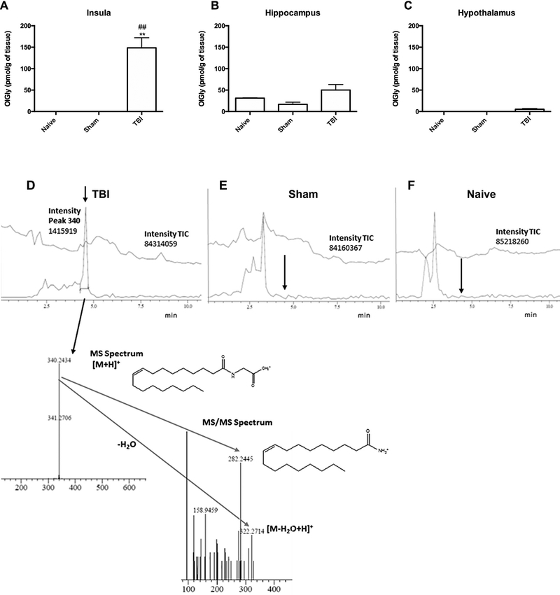
Traumatic brain injury (TBI) leads to increased OlGly levels in the insular cortex of mice. (A) Mice subjected to TBI display significant increases of OlGly in the insula, but not in (B) hippocampus or (C) hypothalamus. Values represent the mean ± SEM of n = 5. **p < 0.001 vs naïve; ##p < 0.001 vs sham. (D) A representative chromatogram shows the presence of OlGly in the insula of mice 24 h following TBI. Injured insula shows formation of OlGly as confirmed by MS and MS/MS spectra. However, OlGly was below the limit of detection in sham (E) and naïve mice (F). The arrow depicts the retention time of OlGly from the standard. The upper chromatogram traces represent the total ion current (TIC), and lower chromatogram traces represent the extracted chromatograms m/z around 340 amu in (D-F).

