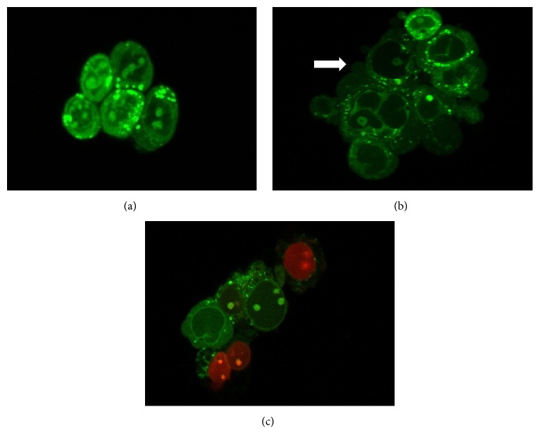
Figure 5.
Fluorescent photomicrographic evidence of HT-29 cells 24 h after treatment. Morphological changes following exposure to treatment are typical of apoptosis; A: viable cell, B: apoptotic cell, C: necrotic cell. The arrow ( ) shows membrane blebbing—one of the characteristics of apoptosis. This was viewed using a laser confocal inverted microscope at magnification of 630X.

