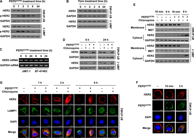
Fig. 2. PEPDG278D, but not Ttzm, induces internalization and lysosomal degradation of HER2 in Ttzm-resistant HER2-BC cells.

(A and B) IB analysis of whole cell lysates from cells treated with PEPDG278D (25 nM, A) or Ttzm (1 μM, B) for 0–24 hours. p-HER2: pY1221/1222-HER2. (C) RT-PCR analysis of gene expression in cells treated with PEPDG278D (25 nM) for 0–24 hours. (D) IB analysis of whole cell lysates from cells treated with vehicle, PEPDG278D (25 nM), and/or chloroquine (25 μM) for 6 or 24 hours. (E) IB analysis of membrane and cytosol fractions from cells treated with vehicle, PEPDG278D (25 nM) and/or chloroquine (25 μM) for 10 min or 6 hours, using MET and GAPDH as loading controls. (F) Confocal fluorescence staining images of HER2, PEPDG278D (staining its His tag) and nuclei (DAPI) in BT-474R2 cells treated with PEPDG278D (25 nM) for 0 or 15 min or 3 hours. Scale bar: 10 μm. (G) Confocal fluorescence staining images of HER2, LAMP1 (lysosome) and nuclei (DAPI) in BT-474R2 cells with or without treatment with PEPDG278D (25 nM) and/or chloroquine (25 μM) for 0–6 hours. Scale bar, 10 μm.
