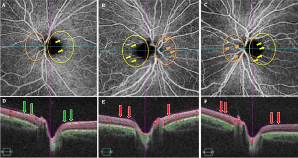
Figure 2.

Shows optical coherence tomography angiography (OCTA) images (A-C) of the superficial vascular networks for the peripapillary nerve fiber layer (pRNFL) in the asymptomatic LHON mtDNA3460 carrier (patient's mother) (A), and affected LHON mtDNA 3460 patient right (B) and left (C) eyes. OCT cross-sections (D-F) show overlaying retinal flow (red) on OCT reflectance (gray scale).
