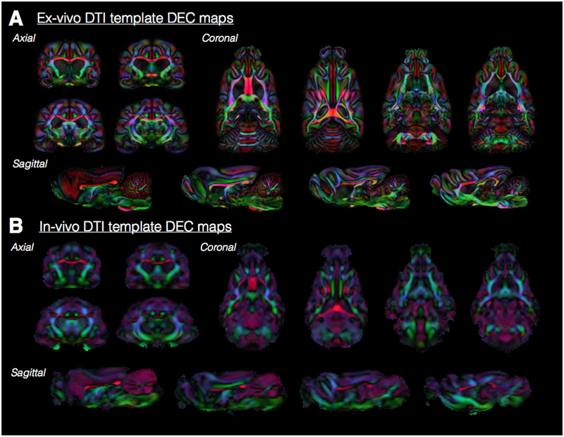
Fig. 6.
Ex-vivo and in-vivo DEC maps throughout the ferret brain. Representative slices from DEC maps for each of the orthogonal orientations are shown to demonstrate the anatomical resolution especially of white matter fiber tracts of the ex-vivo (A) and in-vivo (B) 3D DTI templates. The color coding for primary eigenvector orientations is the same as for Fig. 5.

