
Figure 1.
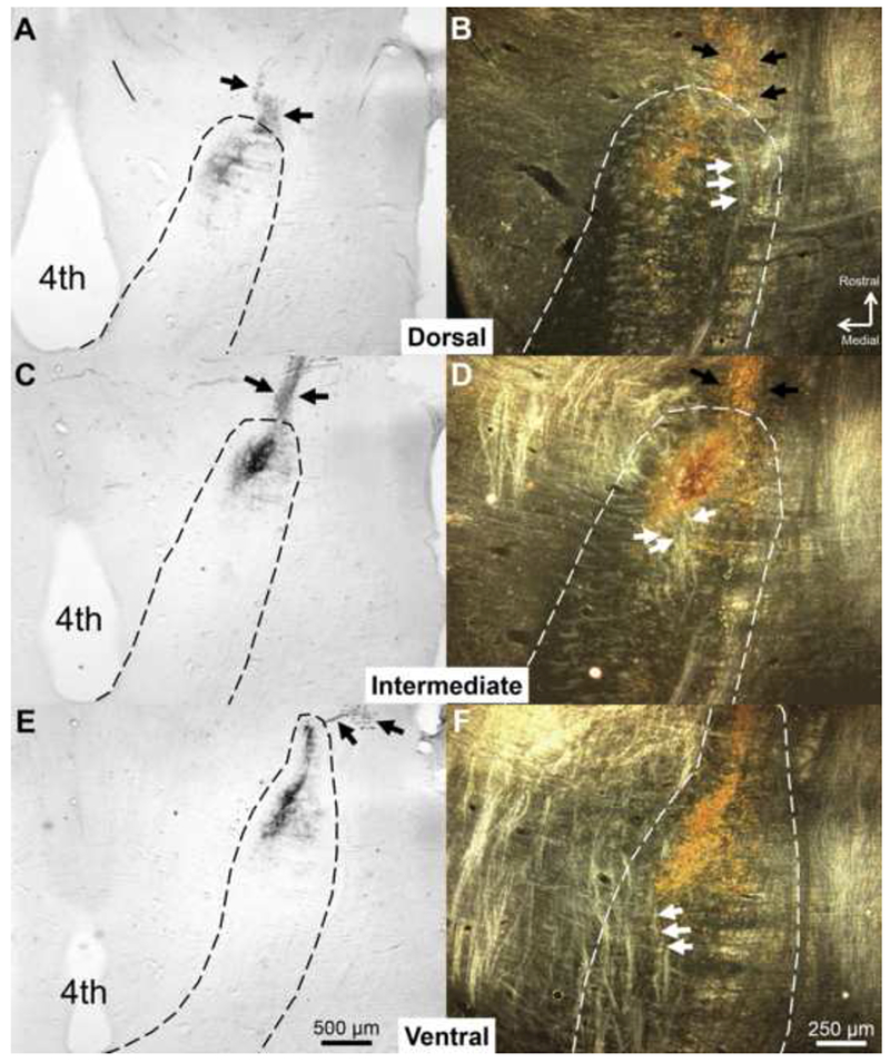
CT terminal field in the dorsal, intermediate, and ventral NTS in a Sham rat labeled with BDA at P60. The NTS was sectioned in the horizontal plane. (A, C, E) Low-magnification, brightfield images of the NTS (dotted line). (B, D, F) Higher magnification images of the NTS are shown using darkfield illumination. In the darkfield images, arrows point to tracts near the CT terminal field that are lighter than surrounding tissue. These tracts are oriented in the rostral-caudal plane and are lateral to gustatory terminal fields in the dorsal NTS. As the NTS progresses ventrally, these tracts situate more medially and move through gustatory terminal fields. We identified sections in which these tracts were within the terminal field area as the intermediate NTS. Black arrows indicate chorda tympani axons projecting toward the NTS. 4th = Fourth ventricle