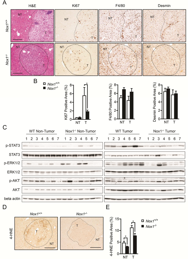
Figure 2. Tumor cell proliferation is reduced in Nox1−/− mice.
(A) Livers were stained with H&E, Ki67 (proliferation marker), F4/80 (marker of macrophages), and Desmin (marker of HSCs). (B) Percentages of positive staining areas of Ki67, F4/80 and Desmin, respectively (n≧6 mice per group). (C) Immunoblot (IB) analysis of p-STAT3, STAT3, p-ERK, ERK, p-AKT, AKT and b-actin in tumor (T) and non-tumor (NT) liver tissues from DEN-injected mice. (D) Representative liver sections that were stained with 4-HNE (4-Hydroxynonenal, marker of ROS). Representative images were taken using objectives x10, Scale bar: 200 μm. (E) Percentages of positive staining areas of 4-HNE (n≧6 mice per group). Data are shown as mean ± s.e.m. Student’s t-test for independent samples and unequal variances was used to assess statistical significance (*P<0.05, **P<0.01, ***<0.001).

