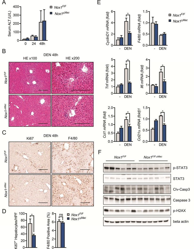
Figure 5. Hepatic inflammation and hepatocyte compensatory proliferation are reduced in Nox1△Mac mice after DEN challenge.
(A) The amounts of alanine transaminase (ALT) were measured in serum 0, 24, 48hrs post DEN challenge (n≧4 mice/group). (B) Representative H&E staining of liver sections 48 hours post DEN injection. (C) Representative immunohistochemical images of murine liver sections 48 hours after DEN injection. Scale bar: 200 μm. (D) Number of Ki67+ hepatocytes per 20x HPF and percentages of positive staining areas of F4/80 in liver sections from indicated mice 48 hrs after DEN injection (n=6 mice/group). (E) Relative expression of CyclinD1, F4/80, Tnf, Il6, Ccl1 and Ccl21⍺ in the total liver extract 48hrs after DEN injection (n=6 mice/group). (F) IB analysis of p-STAT3, STAT3, p-H2AX, cleaved caspase-3 (clv-casp3) and beta actin in the total liver extract 48hrs after DEN injection. Data are shown as mean ± s.e.m.. Student’s t-test for independent samples and unequal variances was used to assess statistical significance (*P<0.05, **P<0.01, ***P<0.0001).

