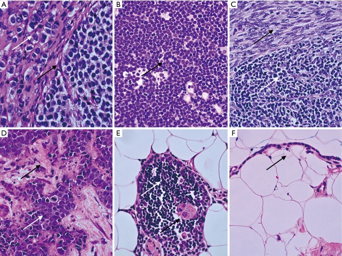
Figure 1.
Representative cases with diagnostic difficulties and with major implications on treatment from the North Estonian Medical Centre (Olympus BX53 equipped with Olympus DP27 camera and Olympus cellSens software). The tissue sections were stained by haematoxylin and eosin (HE) staining (magnification, ×600). (A) Thymic carcinoid tumor. Nuclei with salt-and-pepper chromatin (white arrow) and nested growth pattern of uniform cells (black arrow). Stains for chromogranin A and synaptophysin were positive; (B) T lymphoblastic lymphoma. Destructive growth pattern of monotonous LCA positive lymphoblasts with high mitotic activity (black arrow); (C) AB thymoma. Biphasic tumor with spindle cell epithelial component (black arrow) resembling type A and lymphocyte-rich component resembling type B1 (white arrow); (D) metastatic thymic carcinoma in liver biopsy. Carcinoma cells (white arrow) with high grade atypia, necrosis and central mitosis in the background of desmoplastic stroma (black arrow); (E) thymic tissue with normal lymphocytes (white arrow) and Hassall body (black arrow) surrounded by mediastinal adipose tissue; (F) unilocular cyst lined with thymic cuboid epithelium (black arrow) in mediastinal adipose tissue. LCA, leukocyte common antigen.

