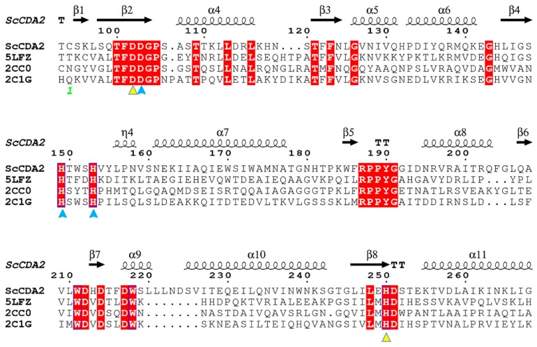
Figure 1.
Structure-based on sequence alignments between four chitin deacetylases (CDAs). The sequence of chitin deacetylase from Saccharomyces cerevisiae (ScCDA2) was aligned with ArCE4A sequences from a marine Arthrobacter species (PDB ID: 5LFZ), the SlCE4 sequence from Streptomyces lividans (PDB ID: 2CC0) and the SpPgdA sequence from Streptococcus pneumoniae (PDB ID: 2C1G). The conserved motifs are highlighted by a red background and the catalytic amino acids are marked with a yellow triangle. Amino acids capable of forming coordinate bonds with Zn2+ are marked with blue triangles. The symbol above the sequence represents the secondary structure, helices represent α-helices, and the arrow represents the beta fold.

