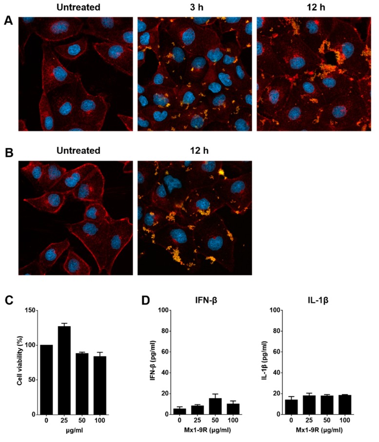
Figure 2.
Transduction of Mx1-9R in vitro. Madin-Darby Canine Kidney (MDCK) cells were incubated with (A) 50 μg/mL or (B) 25 μg/mL of Mx1-9R. At 3–12 h after incubation, cells were permeabilized and stained with anti-Mx1 antibody, then internalization of Mx1 fusion proteins in MDCK cells was detected by confocal microscopy. (C) Cytotoxicity of Mx1-9R was determined by cell viability assay. MDCK cells were incubated with various concentrations of Mx1-9R for 24 h, and cell viability was determined by WST assay (n = 2–3). (D) Mouse bone marrow cells were cultured with Mx1-9R for 18 h and IFN-β and IL-1β levels in culture supernatants were measured by ELISA (n = 3).

