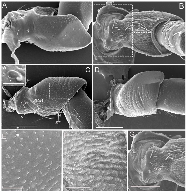
Figure 2.
Micrographs showing the scape of an adult Callitettix versicolor’s (Fabricius) right antenna. (A) Ventral view; (B) Right lateral view; (C) Dorsal side showing sensilla trichodea (ST), sensilla basiconica (SB1) and sensilla campaniformia (SCa1); a. High magnification image of SCa1; (D) Left lateral view; (E) High magnification image of scape surface showing imbricate papillae, indicated by the rectangle given in (C); (F) High magnification image of the surface of concave on the scape, indicated by the small rectangle given in B); (G) High magnification image showing the antenna inserted in antennal foveae on the head capsule, the positions of the images are indicated by the big rectangle given in (B). Scale bars: A, B, C, D = 100 μm; E, F = 25 μm; G = 100 μm; a = 5 μm.

