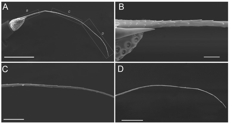
Figure 7.
SEM micrographs showing the flagellum of an adult Callitettix versicolor (Fabricius). (A) Entire flagellum consisting of the bulb-shaped portion and apical arista; (B) High magnification image of the base showing apical arista covered with imbricate papillae; (C) High magnification image of the middle showing apical arista with imbricate papillae decreasing gradually; (D) High magnification image of apical half showing apical arista without imbricate papillae. Positions of images (B–D) are indicated by lettered rectangles given in (A). Scale bars: A = 250 μm; B = 25 μm; C = 50 μm; D = 100 μm.

