Figure 1.
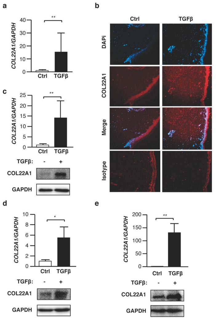
TGFβ increases expression levels of COL22A1 ex vivo and in vitro. (a,b) Human skin samples were treated with TGFβ (10 ng/mL) for 48 and 72 h. (a) Expression levels of COL22A1 were measured in human skin (N = 5); ** p < 0.01. (b) Localization of COL22A1 in ex vivo normal skin. COL22A1 was detected using immunofluorescence in a vehicle control or TGFβ-treated skin tissue. DAPI was used to detect nuclei (original magnification ×40); scale bars = 100 μm. (c) Human normal skin fibroblasts were treated with TGFβ (5 ng/mL) for 24 or 72 h. Expression levels of COL22A1 mRNA were measured in human normal skin fibroblasts (N = 9); ** p < 0.01. Protein levels of COL22A1 in the skin fibroblasts of three healthy donors were analyzed by immunoblotting of the lysates; GAPDH is shown as a loading control. (d) Human normal lung fibroblasts were treated with TGFβ (10 ng/mL) for 48 or 72 h. Expression levels of COL22A1 mRNA were measured in human normal lung fibroblasts treated with a vehicle control or TGFβ (N = 3); * p < 0.05. The protein levels of COL22A1 in lung fibroblasts were analyzed by immunoblotting of the lysates. (e) A549 cells were treated with TGFβ (5 ng/mL) for 24 or 72 h. Expression levels of COL22A1 mRNA were measured in A549 cells treated with a vehicle control or TGFβ (N = 3); ** p < 0.01. Protein levels of COL22A1 in the A549 cells were analyzed by immunoblotting of the lysates.

