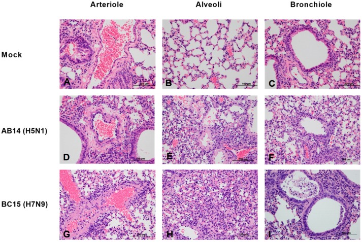
Figure 2.
Lung histopathology of mice after infection with AB14 (H5N1) and BC15 (H7N9) strain isolates. Lung samples were fixed, sectioned, and stained with hematoxylin and eosin. (A–C) Tissues from mock-infected lungs. (D–F) Tissues from mice infected with AB14 (H5N1) showing infiltration of inflammatory cells into the (D) wall of the arteriole, (E) alveolar walls, and (F) bronchiolar epithelium affected by necrosis. (G–I) Tissues from mice infected with BC15 (H7N9) showing necrotic debris and inflammatory cells in the (G) wall of the arteriole, (H) collapsed alveoli, as well as degeneration and necrosis of the (I) bronchiolar epithelium. Scale bar represents 100 µm.

