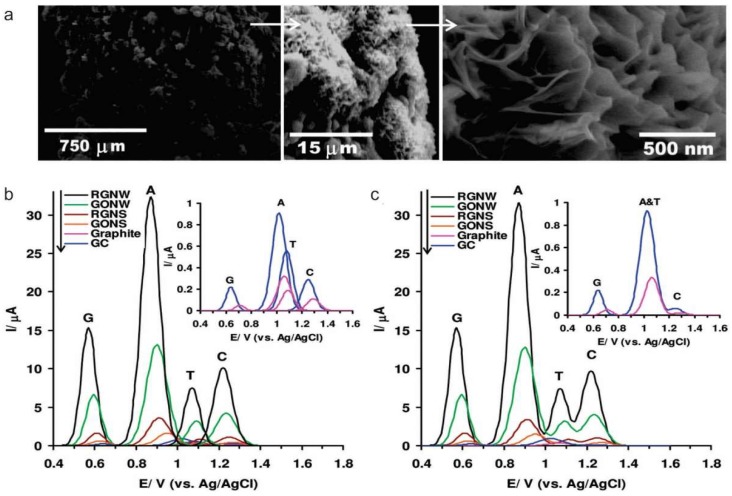
Figure 3.
(a) Scanning electron microscopy images of the graphene oxide nanowalls deposited on a graphite rod by using electrophoretic deposition (b,c) Differential pulse voltammetric profiles of the reduced graphene nanowalls (RGNW), graphene oxide nanowalls (GONW), reduced graphene nanosheet (RGNS), and graphene oxide nanosheet (GONS) electrodes as compared to the graphite and glassy carbon (GC) electrodes for detection of (b) the four free bases of DNA (G, A, T, and C) separately, and (c) equimolar mixture of G, A, T, and C. Reproduced with permission from [71], Copyright American Chemical Society, 2012.

