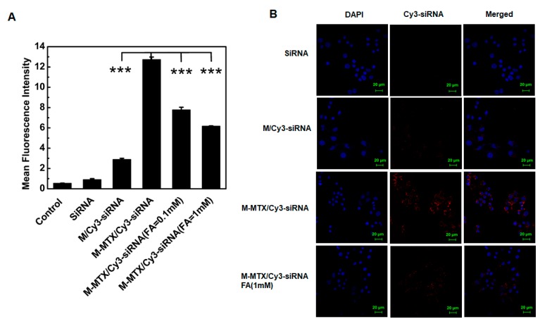
Figure 2.
The cell uptake of M-MTX/Cy3-siRNA in the FR-expressing HeLa cells. (A) Flow cytometry was used to quantify the cellular uptake of different formulations. The HeLa cells were incubated with M-MTX/Cy3-siRNA, M/Cy3-siRNA, and Cy3-siRNA for 4 h at 37 °C. Two concentrations of free folic acid (FA) (0.1 mM and 1 mM) was added 1 h before the M-MTX/Cy3-siRNA (100 nM) was incubated. *** <. (B) The confocal micrographs of HeLa cells were also obtained after incubation with M-MTX/Cy3-siRNA, M/Cy3-siRNA, and Cy3-siRNA for 4 h at 37 °C. Free FA (1 mM) was also added 1 h before the M-MTX/Cy3-siRNA (100 nM) addition. The green bar in the image was 20 μm. The values were the mean ± SEM (n = 3).

