Figure 1.
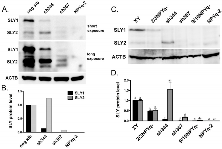
SLY expression in males with NPYq- and Sly-specific deficiency. (A,B) New anti-SLY antibody specifically recognizes SLY1 and SLY2 isoforms. (A) Exemplary Western blot detection of SLY1 (~40 kDa) and SLY2 (~30 kDa) protein in testis lysates from Sly-KD transgenic mice with Sly deficiency (sh344 and sh367). The positive control was a negative sibling of Sly-KD mice (neg sib) while the negative control was a male lacking the NPYq (NPYq-2). (B): Levels of protein expression shown in panel A quantified with ImageJ software (https://imagej.nih.gov/ij/) and normalized with respect to ACTB signal and with neg sib data serving as the normal expression baseline. (C,D) SLY expression in males with NPYq- and Sly-specific deficiency. (C) Exemplary Western blot detection of SLY1 and SLY2 protein in testis lysates from wild-type control (XY), mutant mice with progressively increasing NPYq deficiency (2/3NPYq-, 9/10NPYq-, and NPYq-2) and sh344 and sh367. (D) Levels of protein expression quantified with ImageJ software and normalized with respect to the ACTB signal and with XY data serving as the normal expression baseline. The data represent average ± SEM of several western runs with the following male number per genotype: n = 8, 6, 6, 6, 4, 7 for XY, 2/3NPYq-, sh344, sh367, 9/10NPYq-, and NPYq-2, respectively. Statistical significance (t-test): for each protein isoform, the genotypes marked with different letters are different from each other.

