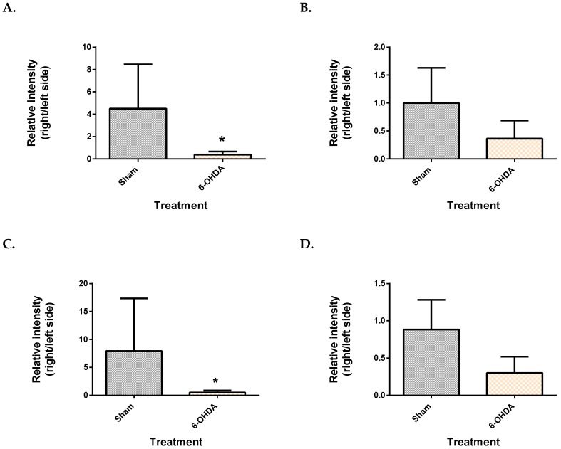
Figure 3.
Comparison of brain metabolite changes between the mesencephalon and cerebellum. Midbrain monopalmitin (A), monostearin (C) myo-inositol (E) were significantly altered while cerebellar monopalmitin (B), monostearin (D), myo-inositol (F) and) were unchanged. Data represent mean ± S.D of at least 5 animals in each group. (* indicates q value < 0.05 using Mann–Whitney test, followed by Benjamini–Hochberg correction).


