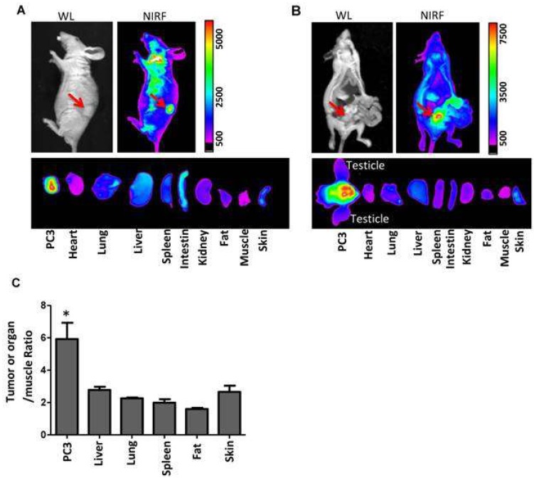
Figure 15.
Imaging-guided drug delivery of NP-AAG in both mouse models bearing (a) subcutaneous and (b) orthotopic PC3 xenograft. In vivo and ex vivo white light (WL) and NIRF imaging of nude mice bearing subcutaneous PC3 xenograft (a) or orthotopic PC3 xenograft (b) 72 h post-injection of NP-AAG. Arrows: tumor site. (c) Analysis of NIRF imaging on each tumor and organs after normalization to the fluorescence of muscle. Reproduced from Reference [52] (Theranostics 06: 1324 image No. 006, open access policy).

