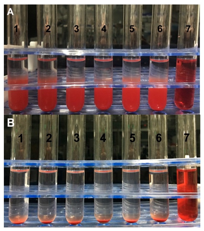
Figure 8.
Photographs of the anti-hemolysis of RBGPs with different concentrations. (A) represents 3 h of incubation at 37 °C, (B) represents 24 h of incubation. The concentrations of RBGPs were 2% (1), 4% (2), 6% (3), 8% (4), and 10% (5). Tube 6 was from the physiological saline group and tube 7 was from the distilled water group.

