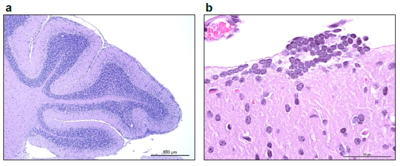
Figure 3.
(a) Low power image taken at 50× total magnification, 5× objective shows multifocal to coalescing nodular remnants of the external granular layer of the cerebellum, with increased cellularity of the molecular layer. (b) Higher magnification image taken at 600× total magnification, 60× objective shows a rest of external granule cells superficial to the molecular layer.

