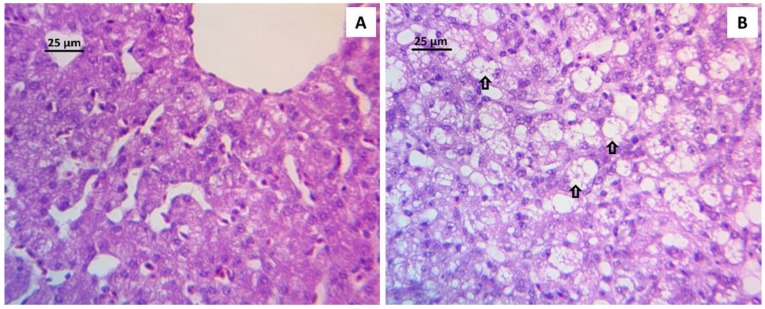
Figure 1.
Liver histopathology showing the different values of the lesion score used to evaluate hepatocellular degeneration. (A) Hepatocellular degeneration score of 0.5: liver section from a bird of NC group showing a scarce number of intracytoplasmic vacuoles. (B) Hepatocellular degeneration score of 3.0: liver section from a bird of AFB1 group, after 21 days receiving a diet with 2 ppm of AFB1, showing an increase of the number of intracytoplasmic vacuoles (arrows). Stain: Hematoxylin and Eosin.

