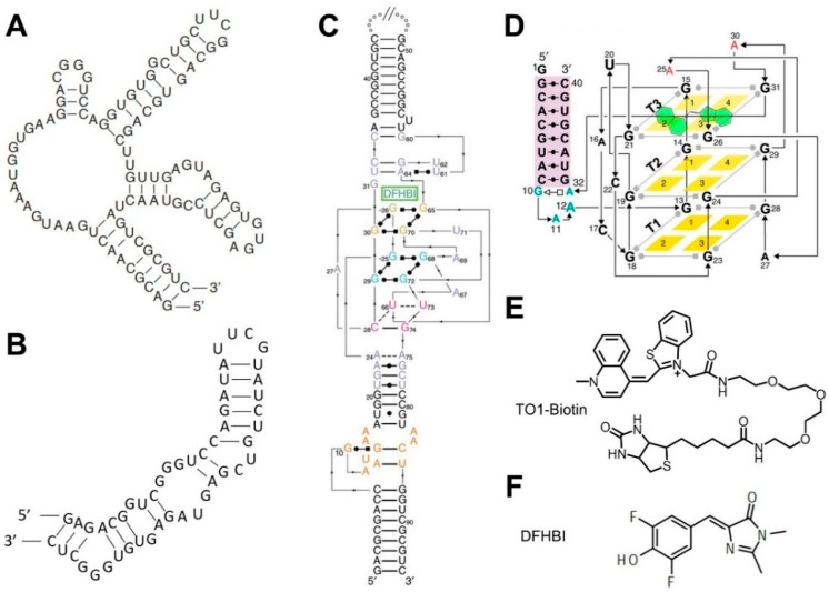
Figure 2.
2D/3D structures of fluorogenic aptamers and the chemical structures of their ligands. (A) Spinach aptamer 2D structure. (B) Broccoli aptamer 2D structure. (C) Spinach aptamer 3D structure [88]. (D) Mango aptamer 3D structure [85]. (E) Mango aptamer’s cognate dye TO1-Biotin. (F) Spinach and Broccoli aptamers’ cognate dye DFHBI.

