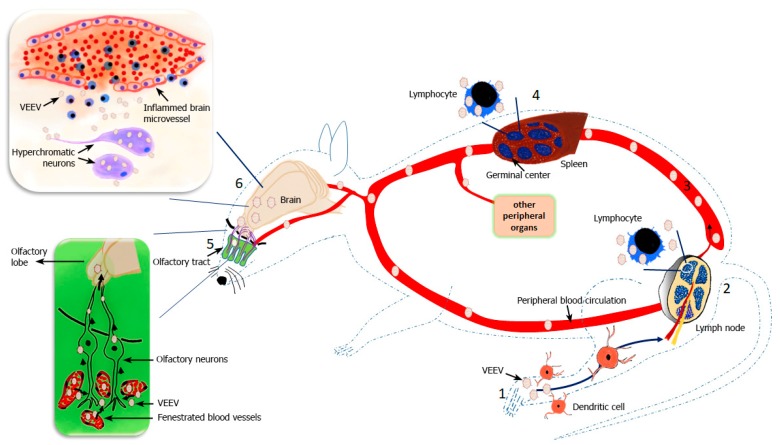
Figure 2.
Biphasic replication of VEEV in mice. Mice infected via VEEV footpad injection mimics the natural mode of transmission by mosquito bites (1). After initial inoculation, regional dendritic cells take up the virus and transport it to the local draining lymph nodes, where VEEV replicates in lymphocytes (2) and is then released into the circulation resulting in viremia (3). VEEV replicates in various tissues, but lymphoid organs such as the spleen are primary replication sites (4). Around 24–36 h post infection, VEEV escapes into the olfactory tract infecting olfactory nerve endings initiating the first step of CNS phase of infection (5). Virus moves through the axons of the olfactory neurons and enters the olfactory lobe of the brain (large insert). In the brain, VEEV primarily infects the neurons, but glia and oligodendrocytes are also targets of VEEV infection. VEEV induced inflammation causes vascular cuffing and alteration in the BBB allowing mononuclear lymphocytes to enter the brain (6). VEEV mediated alteration of the BBB may allow the virus to enter the brain, but this route of entry is debated.

