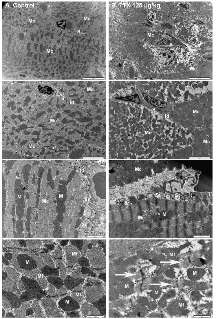
Figure 8.
Electron micrograph of the myocardium from (A) control mice (left panel), and from (B) mice treated daily with 125 µg/kg TTX. Note the different magnifications shown. Scale bar is 10 µm, 5 µm, 2 µm, and 1 µm respectively, from top to bottom panels. Mc: myocardial cells, S: sarcolemma, Id: intercalated disk, Sr: sarcoplasmic reticulum, M: mitochondria, Mf: myofibrils. Evident multifocal vacuolization, sarcoplasmatic dilation, and disintegration of myofibrils (arrows in the bottom right panel) were observed in the heart of TTX-treated mice.

