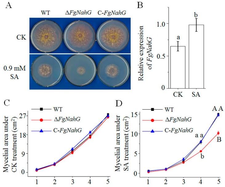
Figure 4.
Effect of FgNahG on mycelial growth. (A) Mycelial growth of the wild-type (WT), ΔFgNahG and C-FgNahG strains on mSNA plates with and without SA at 5 days after inoculations with 1 × 10³ F. graminearum conidia. Five biological replicates were analyzed per treatment. (B) Comparison of FgNahG expression in WT hyphae under the same conditions as the mycelial growth experiment presented in panel (A). (C) Comparison of the mycelial area under CK treatment as panel (A) during 1–5 days after initial inoculation. (D) Comparison of the mycelial area under SA treatment as panel (A) during 1–5 days after initial inoculation. CK, control treatment; SA, salicylic acid treatment. Different lower-case and capital letters above each column indicate significant differences at p ≤ 0.05 and p ≤ 0.01, respectively.

