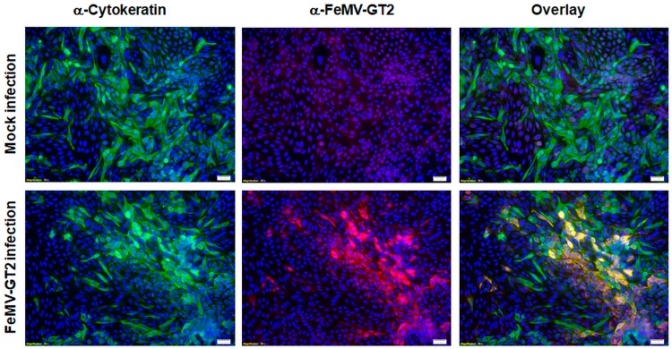
Figure 4.
Susceptibility of primary feline kidney cells to FeMV-GT2. Cells were inoculated with FeMV-GT2-Gordon strain (MOI = 0.5) or mock-infected, fixed five days p.i. and stained for cytokeratin (shown in green) or FeMV-GT2 (shown in red). Double positive cells are orange colored. Cell nuclei were visualized with DAPI (shown in blue). Images were taken at 200-fold magnification. Scale bars represent 20 µm.

