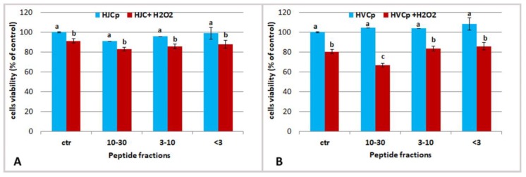
Figure 8.
Effect of oxidative stress on HEKa cells of different concentration of jellyfish (A) and calf skin (B) collagen hydrolyzed peptides MW ranging 30–10 kDa (5 µg/mL), 3–10 kDa (5 µg/mL) and <3 kDa (0.5 µg/mL). Cells viability was valuated 1 h after H2O2 treatment. Data are mean values ± SD of six independent experiments, analysed with ANOVA and Bonferroni post-test (p < 0.05).

