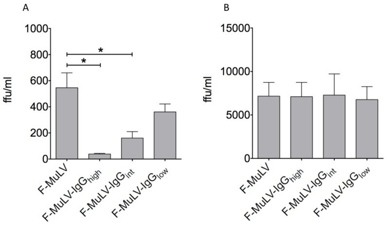
Figure 2.
IgG-opsonization diminishes F-MuLV infection of DCs. F-MuLV stocks were opsonized in the presence of 5 μg/mL (F-MuLV-IgGhigh), 0.5 μg/mL (F-MuLV-IgGint), or 0.05 μg/mL (F-MuLV-IgGlow) FV-specific IgG molecules. (A) DCs or (B) Mus dunni cells were infected with 5000 FFUs of F-MuLV or IgG-opsonized F-MuLV. After overnight incubation, the input virus was removed by washing and cells were further cultivated up to 5 days at 37 °C. Supernatants were collected after 24 h and 5 days of culture and applied in an infectious center assay to determine productive infection. Data represent mean ± SEM of two independent experiments. Data were analyzed by GraphPad PRISM software using ANOVA followed by Dunnet’s multiple comparison test (***, **, * significant at p < 0.001, p < 0.01, p < 0.05, respectively).

