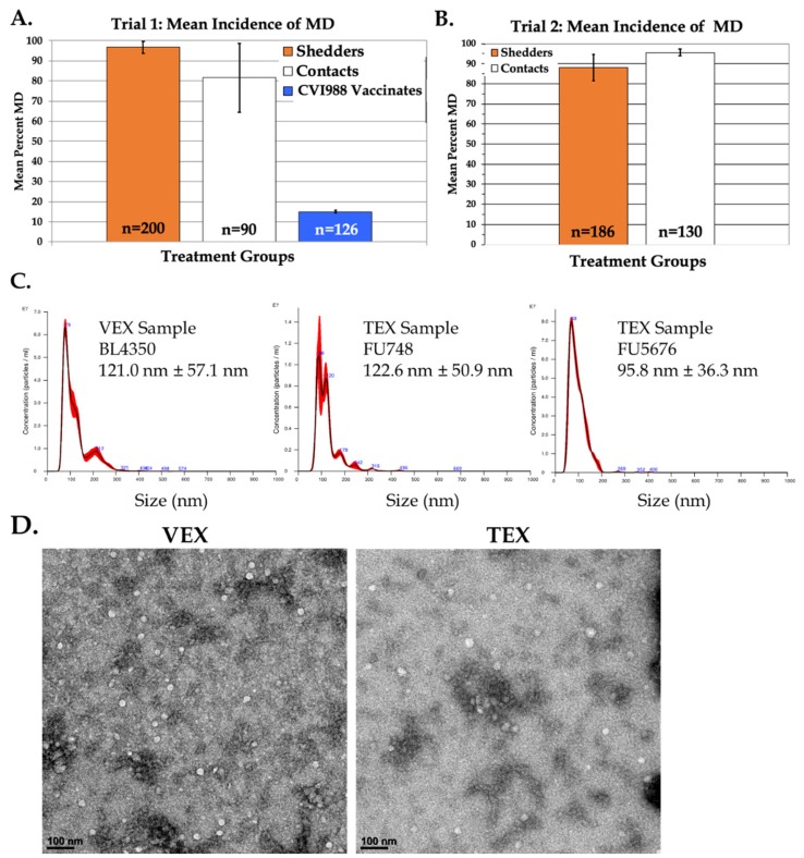
Figure 1.
Exosome source, size, concentration, and ultrastructural characterization. Panels (A,B) show the Marek’s disease (MD) incidences of inoculated shedder (orange), contact-exposed, unvaccinated (white), and CVI988-vaccinated (blue) treatment groups. The numbers of birds for each treatment are given in the bars. Panel (C) shows the nanoparticle size distribution (± SD) and particle concentrations (log particles/mL) of representative VEX and TEX samples. Panel (D) shows representative transmission electron micrographs of Total Exosome Isolation (TEI) reagent-purified VEX and TEX. Vesicles were typically round and varied in diameter from approximately 30–120 nm. Scale bar = 100 nm.

