Figure 3.
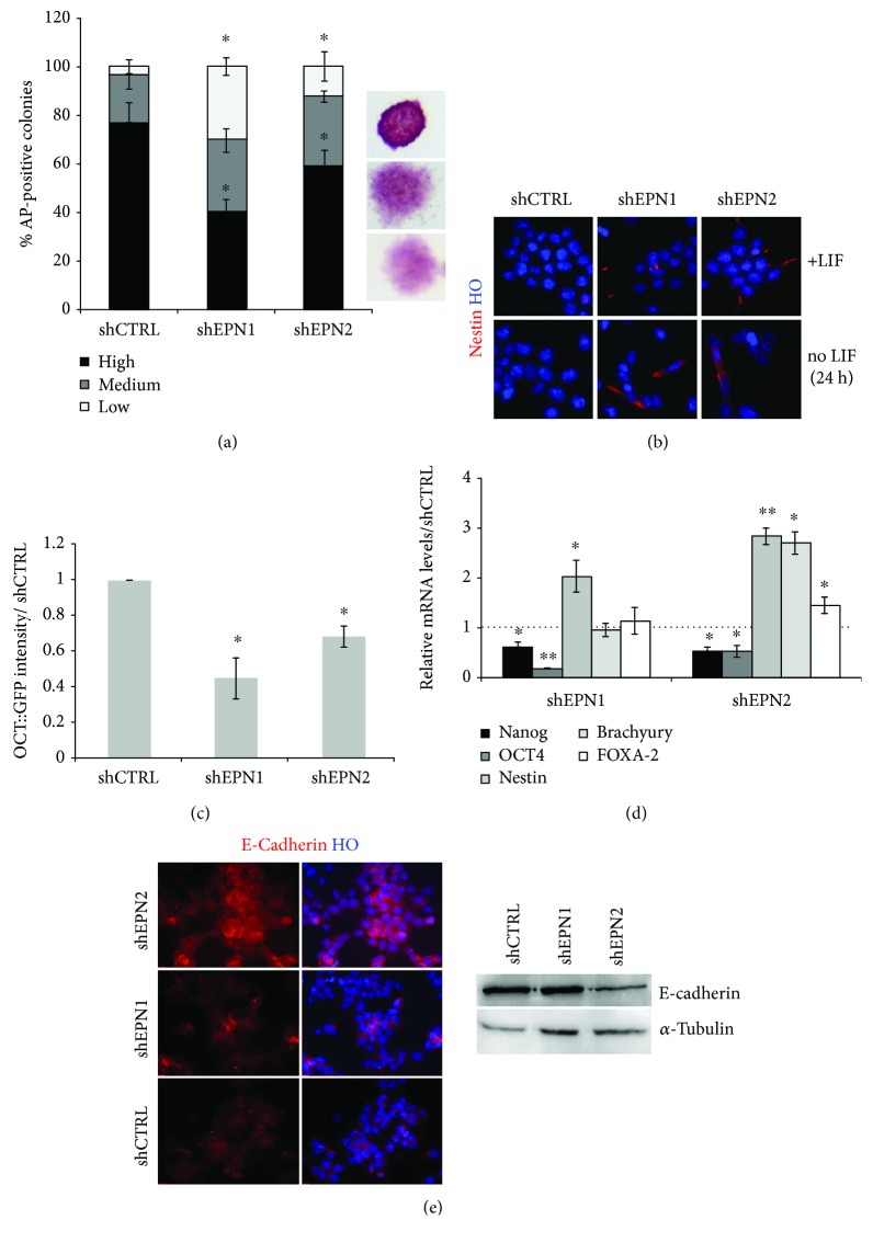
EPN1 and EPN2 KD affects pluripotency and stimulates mESC differentiation. (a) EPN1 and EPN2 silencing in mESCs impairs colony formation capability. Colony formation assays show that EPN1 and EPN2 silencing results in an increase of colonies with low alkaline phosphatase activity and a flat, differentiated morphology. Values are normalized over the control line (shCTRL). (b) EPN1 and EPN2 silencing in mESCs induces nestin upregulation upon LIF withdrawal. Representative immunofluorescence analysis for nestin expression on self-renewing or LIF-deprived EPN1 and EPN2 KD mESCs. Nuclei are counterstained with Hoechst 33258. (c) EPN1 and EPN2 KD results in a significant reduction of OCT4+ve cells after 72 hours of LIF withdrawal. Values are normalized over the control line (shCTRL). (d) qRT-PCR analysis showing relative expression of pluripotency (Nanog and OCT4), neuroectodermal (nestin), mesodermal (Brachyury), and endodermal (FOXA-2) markers in EPNs silenced 46C mESCs. Data are normalized over the control line (shCTRL), using β-actin as the housekeeping gene. (e) Representative immunofluorescence and western blot analyses show the decrease of E-cadherin in EPN1 and EPN2 KD mESCs. α-Tubulin is used as the loading control, and nuclei are counterstained with Hoechst 33258. All data are expressed as the means ± STDV (n = 3 biologically independent experiments). Statistical significance (unpaired t-test): ∗ p < 0.05 and ∗∗ p < 0.001.

