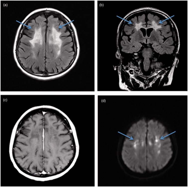
Figure 1.
Brain magnetic resonance imaging demonstrating characteristic findings of adult-onset leukoencephalopathy with axonal spheroids and pigmented glia (ALSP). (a) Fluid-attenuated inversion recovery (FLAIR) image demonstrates subcortical frontoparietal hyperintensities (arrows), with sparing of the temporal lobes and brainstem. (b) Characteristic lack of enhancement on postcontrast T1-weighted axial image (c) and corresponding chronic foci of diffusion restriction (arrows) on diffusion-weighted images (DWIs) (d). The atypical distribution of the non-enhancing white matter disease and persistent chronic ischemic/diffusion restriction changes are unique findings that are highly suspicious for ALSP.

