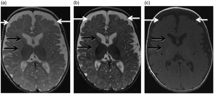
Figure 2.
Brain magnetic resonance imaging (MRI) in patient 2 at age three months. (a) Axial T2 (TR/TE 4350 ms/104 ms), (b) axial inversion recovery (TR/TE 5700 ms/45.8 ms) and (c) axial T1 (TR/TE 500 ms/9 ms) images. There is prominence of the bilateral frontal and temporal subarachnoid spaces indicating frontotemporal brain volume loss, more pronounced than in patient 1 (white arrows). The basal ganglia are small in size and poorly myelinated (black arrows). TR: Repetition Time; TE: Echo Time.

