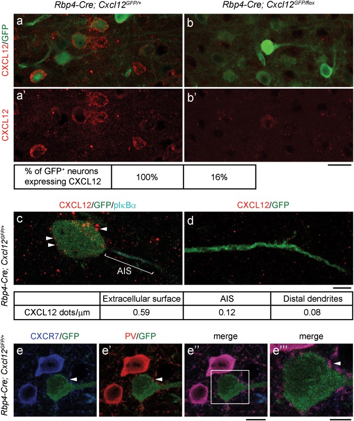
Figure 2.
CXCL12 localizes on the extracellular surface of pyramidal neuron cell bodies, while CXCR7 is present at PV+ interneuron axon terminals in the P45 mPFC. (a, a’, b, and b’) Confocal images show CXCL12 (red) reduction in CXCL12 CKO layer V pyramidal neurons (labeled by GFP). Scale bar, 20 μm. (c–d) Confocal images show that CXCL12 dots (red, indicated by arrowheads) mainly localized on the extracellular surface and inside the cell body (c), while fewer CXCL12 dots were found on the AIS (c, labeled by pIκBα, cyan) and distal dendrites (d, labeled by GFP) of layer V pyramidal neurons. Density of CXCL12 dots represents the number of CXCL12 dots per length of cell body circumference or AIS or distal dendrites. Scale bar, 5 μm. (e–e”) Confocal images show that CXCR7 (blue) localized in the cell bodies and axon terminals of PV+ (red) interneurons. GFP labeled layer V pyramidal neurons encircled by PV+ axon terminals (indicated by arrowheads). Scale bar, 10 μm. (e”‘) The high-magnification image of the region within the white rectangle in (e”). Scale bar, 5 μm.

