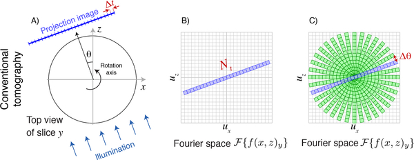
Fig. 1.
Schematic representation of the Crowther criterion in conventional tomography. In conventional tomography, each slice y of the object along the direction of the rotation axis is mapped onto one row of a detector. One then obtains one-dimensional pure projection images of the object with Nt transverse pixels of width Δt in the transverse direction (A), and a depth of precisely one pixel at zero spatial frequency in the axial direction (because there is no way to distinguish between different axial positions in a pure projection). For an angle θ = 0°, the Fourier transform of this image yields an array with Nx = Nt pixels in the transverse or ux direction and Nz = 1 pixels in the axial or uz direction in trasverse-axial Fourier space (B). As the object is rotated, so is the information obtained in Fourier space, so the (ux, uz) Fourier space is filled in as shown in (C). The Crowther criterion of Eq. 3 is effectively a statement that one must provide complete, gap-free coverage of all pixels around the periphery in transverse-axial Fourier space.

