Figure 3.
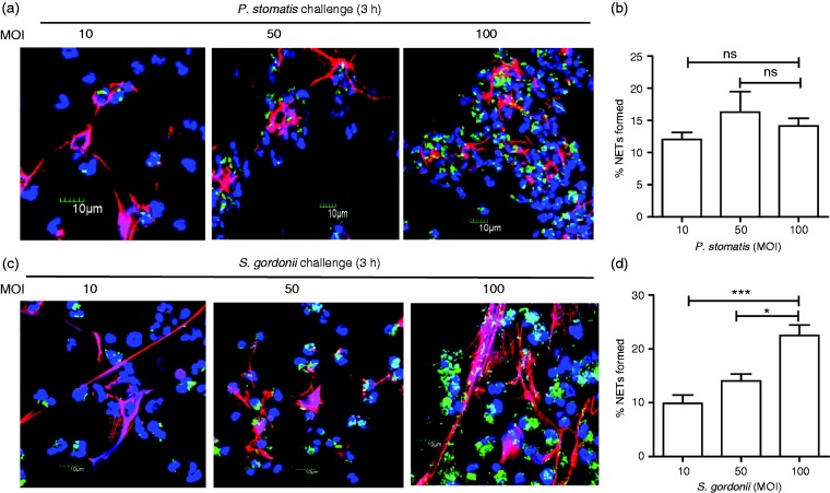
Both P. stomatis and S. gordonii induce NET formation by human neutrophils. Neutrophils were unchallenged (Basal), or challenged with CFSE-labeled P. stomatis (MOI 10, 50, and 100), or CFSE-labeled S. gordonii (MOI 10, 50, and 100) for 180 min. Following infection, cells were fixed and immunostained using Abs directed against MPO (AlexaFluor647), DNA stained with DAPI, and imaged for NET immunofluorescence by confocal microscopy. (a, c) Representative confocal images (from 3 independent experiment of 100 quantified cells per experiment) of CFSE-labeled P. stomatis or S. gordonii challenged neutrophils at 180 min at MOI 10, 50 and 100, respectively. CFSE-P. stomatis or S. gordonii (shown in green); neutrophil nucleus/DNA-DAPI (shown in blue); neutrophil MPO (AlexaFluor647 shown in red); merge image: NET formation. (b, d) Quantification of percentage of NETs formed, by P. stomatis or S. gordonii, using ImageJ. Data are expressed as means of % NETs formed ± SEM from three independent experiments. *P < 0.05, ***P < 0.0001. ns, non-significant.

