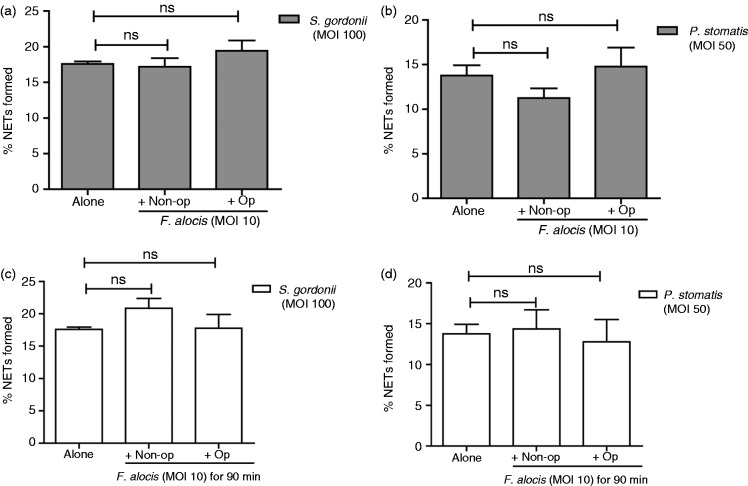
Figure 4.
In co-infection studies, F. alocis has no impact on either inhibition of NET formation or degradataion of pre-formed NETs induced by S. gordonii or P. stomatis. (a) Neutrophils were challenged with HI-labeled S. gordonii (MOI 100, Alone), or co-infected with HI-labeled S. gordonii (MOI 100) + CFSE-labeled non-opsonized F. alocis (MOI 10, Non-op) or CFSE-labeled opsonized F. alocis (MOI 10, Op) for 180 min; or (b) challenged with HI-labeled P. stomatis (MOI 50, Alone), or co-infected with HI-labeled P. stomatis (MOI 50) + CFSE-labeled non-opsonized F. alocis (MOI 10, Non-op) or CFSE-labeled opsonized F. alocis (MOI 10, Op) for 180 min. Following infection, cells were fixed and immunostained using Abs directed against MPO (AlexaFluor647), DNA stained with DAPI, and imaged for NET immunofluorescence by confocal microscopy. (a, b) Quantification of percentage of NETs formed using ImageJ analysis. In (a), data are expressed as means of % NETs ± SEM from four independent experiments. In (b), data are means ± SEM from three independent experiments. ns, non-significant. (c) Neutrophils were challenged with HI-labeled S. gordonii (MOI 100, Alone) for 180 min, or HI-labeled S. gordonii (MOI 100) for 90 min and then infected with CFSE-labeled non-opsonized F. alocis (MOI 10, Non-op) or CFSE-labeled opsonized F. alocis (MOI 10, Op) for additional 90 min or (d) challenged with HI-labeled P. stomatis (MOI 50, Alone) for 180 min, or HI-labeled P. stomatis (MOI 50) for 90 min and then infected with CFSE-labeled non-opsonized F. alocis (MOI 10, Non-op) or CFSE-labeled opsonized F. alocis (MOI 10, Op) for additional 90 min. Following infection, cells were fixed and immunostained using Abs directed against MPO (AlexaFluor647), DNA stained with DAPI, and imaged for NET immunofluorescence by confocal microscopy. (c, d) Quantification of percentage of NETs formed using ImageJ analysis. Data are expressed as means of % NETs ± SEM from three independent experiments. ns, non-significant.

