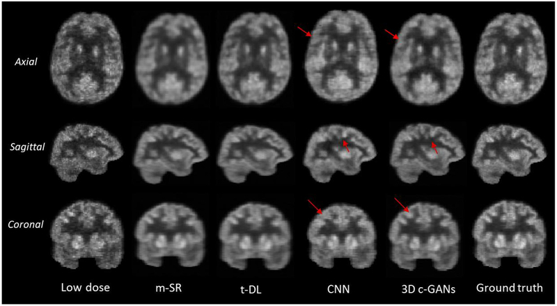
Fig. 9.
Qualitative comparison of low-dose PET images, estimated by the mapping based sparse presentation method (m-SR), by semi-supervised tripled dictionary learning method (t-DL), by convolutional neural networks (CNN), and by the proposed concatenated 3D c-GANs method (3D c-GANs), as well as the real full-dose PET images (Ground truth). In the axial and coronal images, the left side of the image is the right side of the brain, and the right side of the image is the left side of the brain.

