. 2015 Dec 17;1(3):180–189.

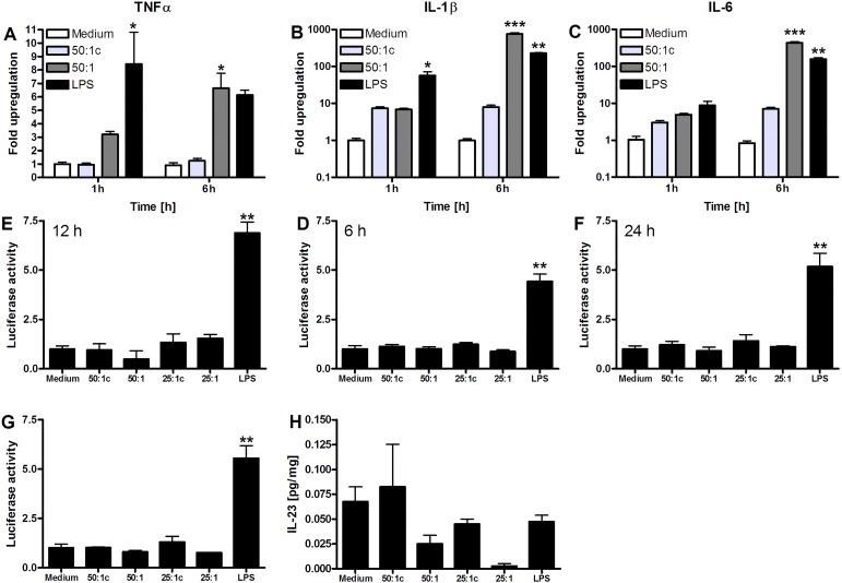
Figure 2. Pro-inflammatory signaling by DAMP-exposed RAW 264.7 macrophages. A: TNFα, B: IL-1β, and C: IL-6 mRNA expression after 1-h and 6-h DAMP incubation. All results are presented as fold upregulation compared to medium incubation (N = 3-4 per group). D-G: Luciferase reporter assay of RAW 264.7 NF-κB/LUCPorter cells following medium-, DAMP-, or LPS stimulation after D: 6 h, E: 12 h, and F: 24 h (N = 3 per group). G: Luciferase reporter assay after stimulation with DAMPs derived from ischemia-subjected necrotic cells, medium, or LPS after 24 h of exposure (N = 3 per group). Luciferase activity is expressed as the fold change relative to control. H: IL-23 production by murine macrophages in response to AML12 hepatocyte-derived DAMPs measured by ELISA in RAW 264.7 cell supernatant and corrected for protein (N = 4 per group). All data represent mean ± SEM. * indicates p < 0.05, ** indicates p < 0.01, *** indicates p < 0.001 compared to the medium samples.

Figure 2.