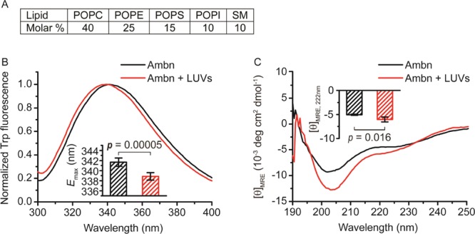
Figure 1.

Changes in Ambn secondary structure as the result of LUV addition. (A) Lipid composition used to assemble 100 nm diameter LUVs (see experimental). (B) Fluorescence and (C) CD spectra of Ambn with 300 μM 100 nm LUVs. The inset in (B) shows the emission maxima of the Trp fluorescence spectra (Student’s t-test, n = 6). The inset in (C) shows the changes of averaged mean residue ellipticity values at 222 nm ([θ]MRE,222nm) in the presence of LUVs (Student’s t-test, n = 4).
