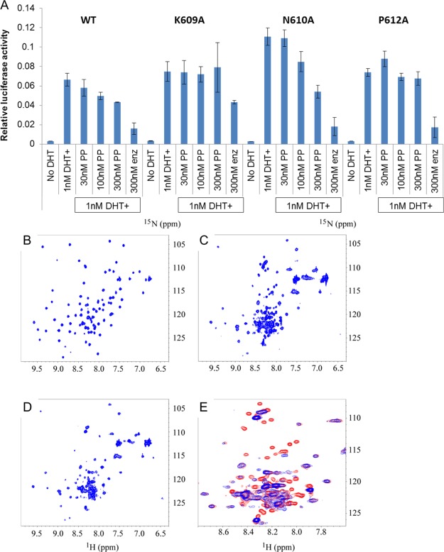
Figure 1.
Interaction with the DBD. (A) Alanine mutations of AR at K609, N610, and P612 were transfected into PC3 prostate cancer cells, along with PSA-luciferase and SV40-renilla luciferase control reporter plasmids. Following overnight treatment with the indicated drugs, the luciferase activity was quantified in quadruplicate samples. PP inhibits the activity of wild type and N610A AR but not K609A or P612A AR. (B–E) 1H–15N HSQC spectra of 15N-labeled AR DBD and its complex with the DNA duplex in the absence and presence of P24. (B) HSQC spectrum of 15N-labeled AR DBD. (C) HSQC spectrum of 15N-labeled AR DBD in complex with DNA. (D) HSQC of the 15N-labeled AR DBD and DNA complex in presence of P24. (E) Zoomed-in region of overlay from the spectra of C (in red) and D (in blue) to show the difference caused by the addition of P24 to the protein–DNA complex.

