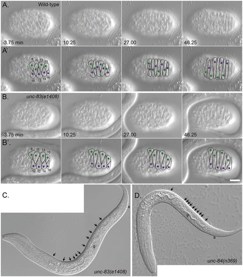
Fig. 1.
Nuclear migration in embryonic hyp7 precursors. (A, B) DIC images from a time-lapse series of images of nuclear migration in dorsal hyp7 precursors in wild-type (A) and unc-83(null) (B) embryos. The time that cell 12 completed intercalation was defined as t = 0. Dorsal view, anterior is to the left. (A’, B’) Cell borders are outlined in black, nuclei migrating left to right are purple, and nuclei migrating right to left are green. Bar, 10 μm. Reproduced from [15] with permission from The Journal of Cell Biology. (C, D) L1 larva showing hyp7 nuclei mislocalized in the dorsal cord (arrowheads) in unc-83(null) (C) and unc-84(null) (D). In wild type, there would be no nuclei in the dorsal cord. Not all nuclei are seen in this focal plane. Lateral view; anterior is to the left, and dorsal is up. The four-cell germ line (g) and the anus (a) are marked to help with dorsal-ventral orientation. Note that the anchorage phenotype shown in (D) is the most severe phenotype seen in unc-84(null) larvae

