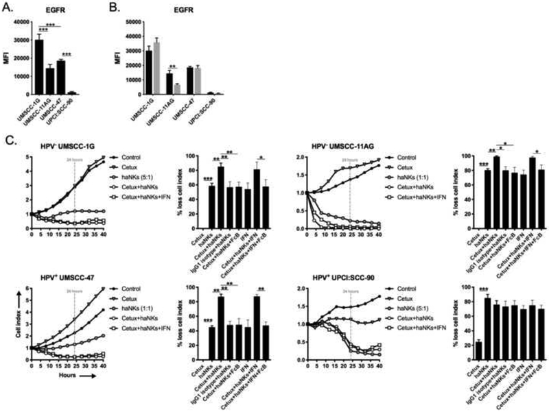
Figure 5 – Cetuximab variably enhances haNK cytolysis of HNSCC cell lines via ADCC.
Baseline (A) and IFNγ-inducible (B) expression of cell surface EGFR in HNSCC cells assessed by flow cytometry. (C) HNSCC lines were plated in the presence or absence of cetuximab (1 μg/mL), with or without IFNγ, and allowed to gain impedance overnight prior to the addition of haNKs at the indicated E:T ratios. Cell index plots normalized to the addition of haNKs at time 0. For each model, representative impedance plots for select conditions shown on the left, and percent loss of cell index relative to control (no haNKs) 24 hours after the addition of haNKs quantified on the right. IgG1 isotype control antibody and CD16 (FcR) blocking antibodies used as controls. Representative results from one of at least two independent assays shown, each performed in at least technical triplicate. *, p<0.05; **, p<0.01; ***, p<0.001; student’s t-test or ANOVA.

