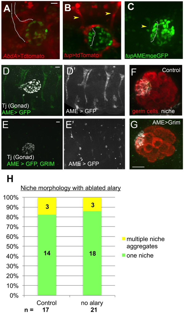
Figure 3: An alary muscle is located near the niche.
A) Live-imaging AbdA>tdTomato revealed a Y-shaped structure anterior to assembled hub (dotted line, six4-nlsGFP). B) Tup> tdTomato identified the structure as an alary muscle (AM); one located at the anterior and one at the posterior of the gonad (arrowheads). C) tupAME-moe::GFP specifically marked the AM (arrowhead), which was present as pro-hub cells assembled (outline, six4-nlsGFP). (D, D’) Control embryo carrying tupAME-GAL4>GFP, stained with Traffic jam to mark the gonad. D) A control embryo with GFP under control of the AME driver, revealing the position of the alary muscles. E, E’) Embryo with Alary muscles ablated by tupAME-GAL4>Grim, GFP. Lone GFP+ cells are likely pycnotic, unfused alary muscle precursors. Dissected gonads from control (F) and alary ablated (G) embryos, stained for germ cells (Vasa, red) and hub cells (FIII, white). Ablation did not disrupt niche formation, which was consistently at the gonad anterior, located opposite the msSGPs (not shown in the image). (H) Quantification of niche morphology from control and ablated embryos. Scale bars are 10 microns.

