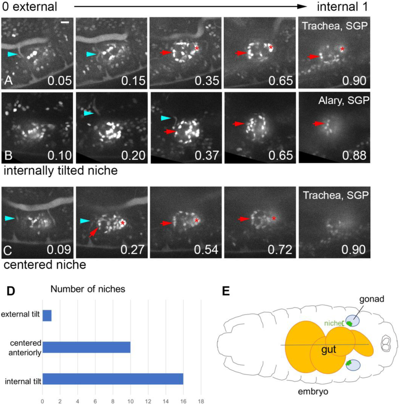
Figure 4: The hub often adopts an internal tilt, relative to A-P axis of embryo.
A, B, C)The final time point from a movie visualizing niche assembly (six4-nlsGFP). Each sequence shows Z slices starting from the external region of the gonad, and moving more internally, with the fractional position along the external-internal gonad axis indicated (0, most external; 1, most internal). Niche cells (arrow), msSGPs (asterisk), and either trachea (A, C; breathless>GFP, blue arrowhead), or the Alary (C; tupAME-GFP, blue arrowhead). Scale bar is 10 microns. D) Frequency distribution of the tilt of the niche. E) Schematic of an embryo showing a dorsal view of the gut, the gonads, and an internally tilted niche in each gonad.

