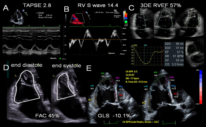
Figure 2.
Right ventricular function parameters measured by echocardiography. (A) Tricuspid annular plane systolic excursion, TAPSE. (B) Tissue Doppler imaging at the RV base demonstrating regional longitudinal shortening, S wave. (C) 3D echo assessment of RV volumes and ejection fraction (RVEF). (D) Fractional area change calculation from the RV focused four-chamber view. The endocardium is traced (beneath the trabeculations) in end-systole and end-diastole. (E) Lateral and septal wall strain (six-segment) imaging to calculate global longitudinal strain, GLS, of the RV. RV strain is reduced −10.1%.

 This work is licensed under a