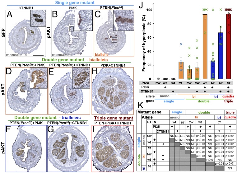
Fig. 1.
Genotype-dependent uterine histopathology at 2 mo after in vivo mutagenesis. Immunohistochemical analyses for green fluorescent protein (GFP) (markers for Cre activity) (A) and pAKT (B–I) highlighted the mutant epithelial cells in the uteri from mice carrying indicated mutation(s). (Scale bar: 500 μm.) (J and K) Frequency of hyperplastic lesions in mice with different genotypes. (J) Result is presented by average ± SE of all mice in each group including ones negative for epithelial lesions. (K) Summary of statistical analysis by one-way ANOVA with post hoc Tukey’s HSD test.

