Fig. 1.
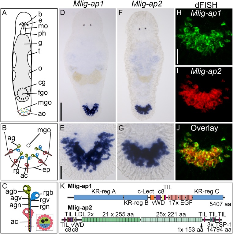
Location, expression, and organization of M. lignano adhesive proteins. (A–C) Schematic drawing of adult M. lignano with detailed structure of adhesive organs. (D–J) Expression of Mlig-ap1 and Mlig-ap2 mRNA visualized with colorimetric WISH (D–G) and dFISH (H–J). Note the coexpression in the same cells of both mRNAs in the overlay. (K) Schematic drawing of the protein structure of the two adhesive proteins. Protein domains: aa, amino acids; c8, domain of eight conserved cysteines; c-Lect, c-type lectin domain; EGF, epidermal growth factor-like domain; LDL, low-density lipoprotein receptor-like domain; TIL, trypsin inhibitor-like domain; TSP-1, thrombospondin 1-like domain; vWD, von Willebrand factor type D-like domain. ac, anchor cell; ag, adhesive gland; agb, adhesive gland body; agn, adhesive gland neck; agv, adhesive gland vesicle; ao, adhesive organs; b, brain; cg, cement glands; e, eyes; eg, egg; ep, epidermis; fgo, female genital opening; g, gut; mgo, male genital opening; mo, mouth; o, ovaries; ph, pharynx; rg, releasing gland; rgb, releasing gland body; rgn, releasing gland neck; rgv, releasing gland vesicles; t, testes; tp, tail plate. [Scale bars, 100 µm (D) and 20 µm (E and H).] Schemes modified from ref. 29, which is licensed under CC BY 4.0.

