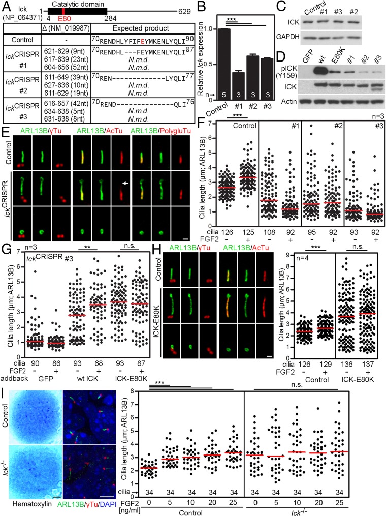
Fig. 7.
FGF increases cilia length via ICK. (A) CRISPR/Cas9 targeting of Ick in triploid NIH 3T3 cells. IckCRISPR #1–3, three clones with all three alleles targeted; the positions of deletions are indicated (N.m.d., nonsense mediated decay). (B) Ick expression was analyzed by qPCR, and normalized to Gapdh. (C) ICK protein levels were analyzed by Western blot. (D) 293T cells were transfected with FLAG-tagged wild-type ICK, kinase-dead ICK-E80K or ICK variants harboring in-frame deletions as shown in A, and Western blot for ICK activating phosphorylation (p) at Y159 was used to determine the kinase activity. (E) Cilia in IckCRISPR cells were visualized by ARL13B, acetylated tubulin (AcTu), polyglutamylated tubulin (PolygluTu) or γ-tubulin (γTu) immunostaining. Both long and extremely short cilia signals are shown for IckCRISPR cells. (Scale bar, 1 µm.) Missing AcTu staining is indicated (arrow). (F) IckCRISPR cells were treated with FGF2 for 12 h and the cilia length was measured; black dots, individual cilia; red bars, medians. (G) IckCRISPR #3 cells were transfected with wt ICK or ICK-E80K, treated with FGF2, and the cilia length was measured. GFP transfection was used as a control. FGF2 extended primary cilia length in wild-type ICK add-back cells but not in ICK-E80K cells. (H) Human control and ICK-E80K fibroblasts were serum starved and immunostained to visualize cilia. (Scale bar, 1 µm.) Cells were treated with FGF2 and the cilia length was measured. (I) Micromasses produced from limb buds of either Ick+/+ (control) or Ick−/− E12 mouse littermates were treated with FGF2 for 24 h and stained with hematoxylin or ARL13B/γ-tubulin. [Scale bars: 1 mm (Left) and 5 µm (Right).] The cilia lengths were measured and graphed. Student’s t test, **P < 0.01, ***P < 0.001; n.s., nonsignificant.

